The Microscopy and Imaging Facility Center
Microscope is crucial to cell biology researches. With the development of physics, optics, computer science and manufactory technology, microscope has developed greatly. Cutting-edge Scientific achievements can not be made without microscopes.
In order to adapt to the development of science and technology, The Microscope and Imaging Facility Center has been established in the institute of biomedical science in 2008. Our goal is to provide the high-efficiency and high-quality microimaging service to researchers.
Now we have several Olympus inverted microscope. an Olympus stereomicroscope, a leica sp5 Confocal laser scanning microscopy, several anatomize lens, a leica Fluorescence Upright Microscope.
Director:Wang ping
Technician:Pan weijuan, Liu dongmei
Equipment and main uses
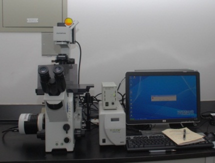
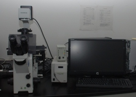
IX71 inverted microscope. The IX71 inverted research microscope is designed to accommodate a wide range of advanced research techniques. The IX71's modular frame and optical design provides 9 access ports for multiple input or output devices. Up to four ports can have simultaneous access to a primary image. The IX71 system can easily accommodate multi-wavelength, advanced fluorescence techniques.
.
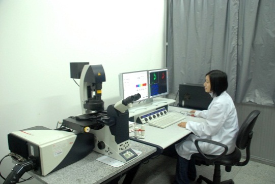
leica sp5 Confocal laser scanning microscopy The Leica TCS SP5 Confocal fully covers a broad range of requirements in confocal and multiphoton imaging with excellent overall performance. The system provides the full range of scan speeds at the highest resolution.
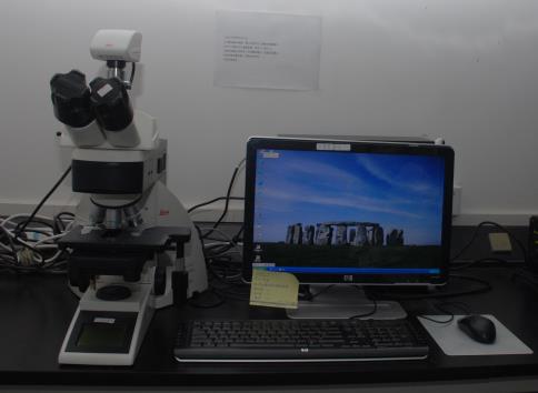
leica Fluorescence Upright Microscope
IX71 inverted microscope can provide single reflection, high intensity, highly corrected images including transwell wound healing angiogenesis.
IX71 living cell culture system can be used to record cell culturing, and then to observe cellular morphologic change and migration and so on.
It can be used for Immunofluorescence observation and photography, such as protein location, virus screening and computing efficiency of virus infection, etc.
Upright Microscope delivers high performance automated microscopy for research applications and provide an exceptionally stable specimen surface for greater reproducibility in imaging experiments.
The Olympus stereomicroscope series is designed to increase contrast and resolution for obtaining the highest image quality during macro observation at low magnification, as well as optimum resolution at high magnification. Enhanced versatility is assured by high zoom ranges and increased focal depth. The new features, automated options and eco-friendly lighting allow for high level application-oriented imaging and digital archiving tasks
The Leica TCS SP5 Confocal fully covers a broad range of requirements in confocal and multiphoton imaging with excellent overall performance. The system provides the full range of scan speeds at the highest resolution.
Management and operation
To avoid the damage caused by false operation, operational processes should be obeyed when operating microscopy. Everyone who’s not familiar to operating microscopy should undergo some training before using the device.
Confocal laser scanning microscopy
Operating Confocal laser scanning microscopy correctly and skillfully is not only the premise of high-quality image and realized experimental purpose but also guarantee the lifespan of the device.
To guarantee the device work better, we set the following rules.
Specific person should be responsible for the microscope and the user should make a reservation beforehand.
Everyone should log on to the computer connected to the microscope with their real name.
Reservations should be made before using the device. Everyone can use the device no more than four hours once. To extend the lifespan of the device, frequent start should be avoided. Seeing that staff can adjust the reservation to guaranteeing the proper use of device, everyone using the device should confirm with the staff before using it.
To enhance the service efficiency, the confocal microscopy is not available to experiments that can be conducted by general microscopies. In addition, immobilized cells and tissue samples can be photographed by the device only if they have been observed by general ones.
Please discuss the experimental designs and make an appointment with the staff beforehand if your photography requests for long time or other special requirements.